In the realm of ear well being, correct analysis is essential for efficient therapy, particularly when coping with situations that may result in listening to loss. Traditionally, otolaryngologists have relied on the otoscope, a tool that gives a restricted view of the eardrum’s floor. This standard instrument, whereas helpful, has its limitations, notably when the tympanic membrane (TM) is opaque resulting from illness.
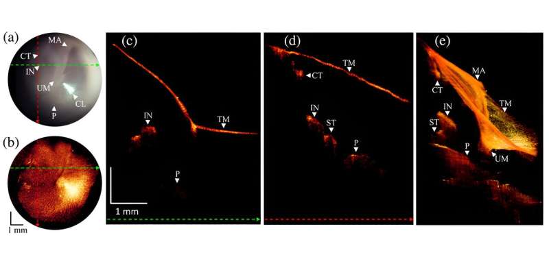
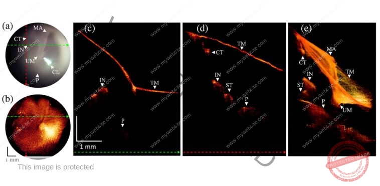
Enter a groundbreaking development from the University of Southern California’s Caruso Department of Otolaryngology: a conveyable OCT otoscope that integrates optical coherence tomography (OCT) with the normal otoscope, to enhance diagnostic capabilities in listening to clinics. As reported in Journal of Biomedical Optics, the built-in system permits clinicians to acquire detailed views of each the floor and the deeper constructions of the eardrum and center ear, enabling a extra complete image of ear well being and enhancing diagnostic accuracy.
Traditional otoscopes solely permit for a superficial examination of the TM, usually lacking deeper pathologies. In distinction, the OCT otoscope combines the acquainted otoscopic view with high-resolution imaging of the inside constructions of the TM and center ear (ME), providing a clearer and extra complete view, which will help in diagnosing situations that have been beforehand missed.
This state-of-the-art system contains a 7.4 mm area of view and spectacular lateral and axial resolutions of 38 micrometers and 33.4 micrometers, respectively. It additionally integrates superior algorithms to boost picture readability and proper distortions, making certain exact and dependable outcomes.
During a scientific examine at USC Keck Hospital, the researchers examined the OCT otoscope on over 100 sufferers. These assessments exhibit the brand new system’s skill to disclose pathological options that have been beforehand invisible utilizing normal otoscopy.
Notably, the article showcases just a few scientific purposes together with monitoring myringitis, tympanic membrane perforation therapeutic, retraction pockets, and subsurface scarring / air pockets; the brand new imaging system recognized a number of vital situations that weren’t obvious by way of conventional strategies, providing useful insights for simpler administration and therapy of ear ailments.
The OCT otoscope’s design permits for seamless integration into current scientific workflows, with an easy-to-use interface managed by a foot pedal for picture acquisition. This user-friendly strategy ensures that the system may be readily adopted by clinicians, offering them with a strong new instrument for diagnosing and managing TM and ME issues.
Overall, this development marks a major step ahead in otolaryngology, enhancing the precision of ear examinations and probably main to raised outcomes for sufferers affected by listening to loss resulting from ear pathologies. As this know-how turns into extra broadly accessible, it guarantees to rework the best way ear well being is assessed and handled, providing hope for extra correct diagnoses and improved affected person care.
More data:
Wihan Kim et al, Optical coherence tomography otoscope for imaging of tympanic membrane and center ear pathology, Journal of Biomedical Optics (2024). DOI: 10.1117/1.JBO.29.8.086005
Citation:
New imaging system improves ear illness analysis (2024, August 23)
retrieved 24 August 2024
from
This doc is topic to copyright. Apart from any honest dealing for the aim of personal examine or analysis, no
half could also be reproduced with out the written permission. The content material is supplied for data functions solely.


